Ultrasound-guided central venous catheterization model

【Superior Ultrasound Imaging for Guided Training】
Engineered with ultrasound-optimized tissue that delivers exceptional acoustic transparency. The internal jugular vein, carotid artery, trachea, and clavicle are clearly distinguishable under real ultrasound devices – supports both ultrasound-guided and landmark-based blind approaches.

Related Products

Ultrasound-Guided Radial Artery Puncture Training Model

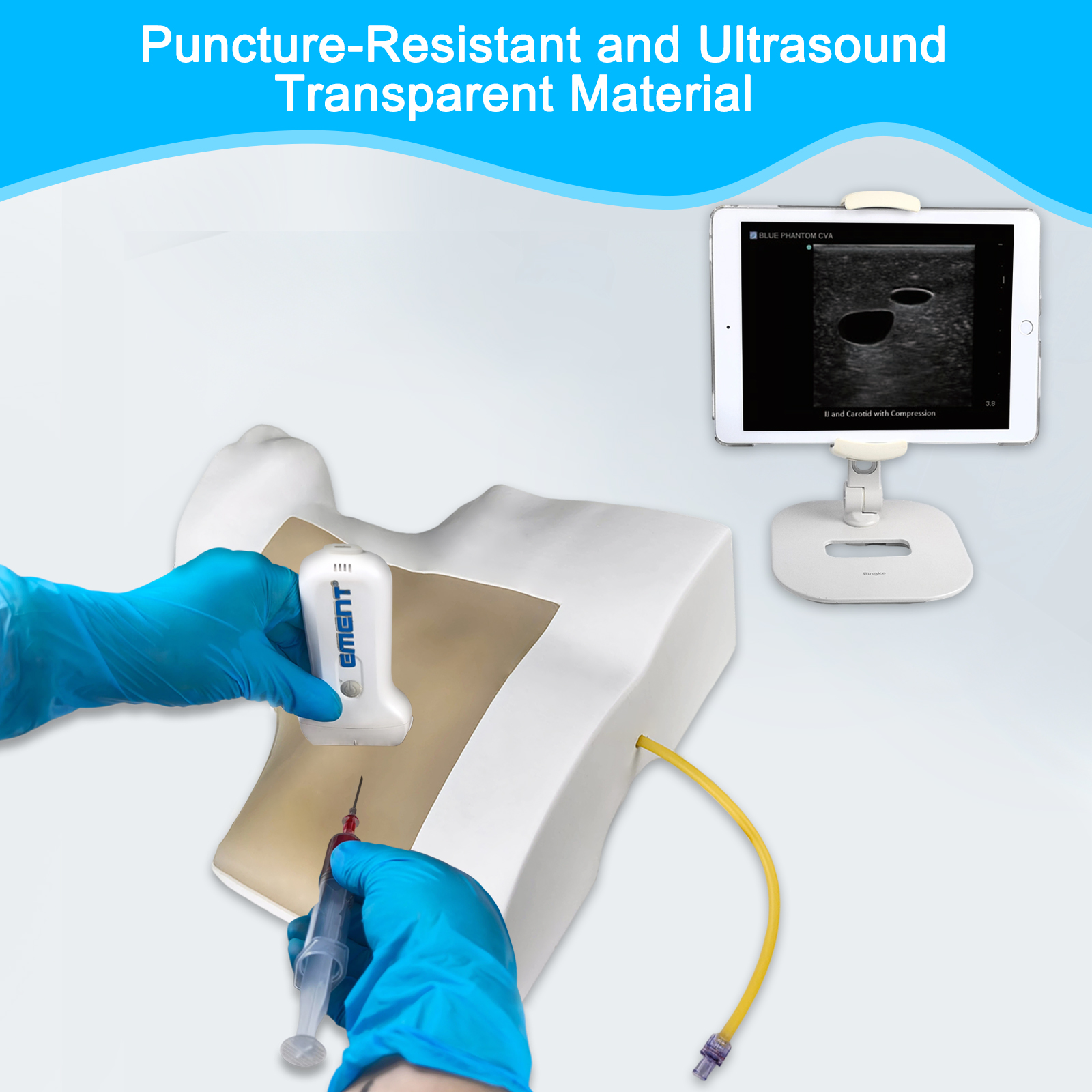
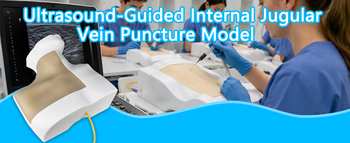
1. High-precision biomimetic anatomical structure for clear and lifelike ultrasound imaging 
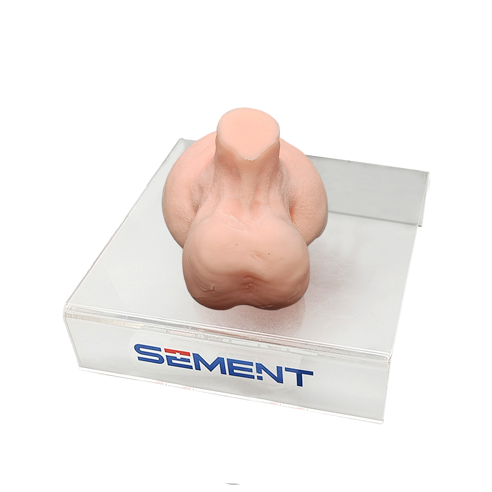
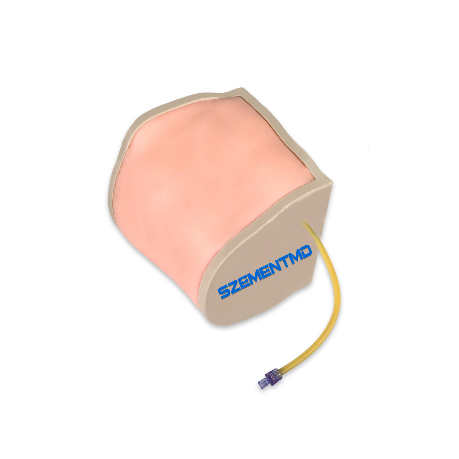
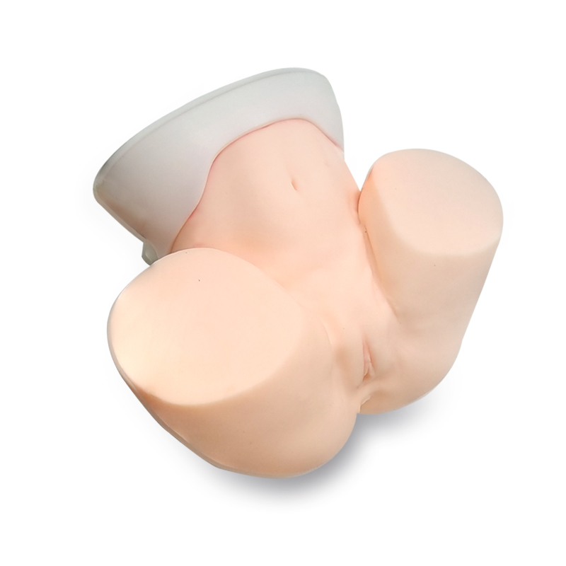
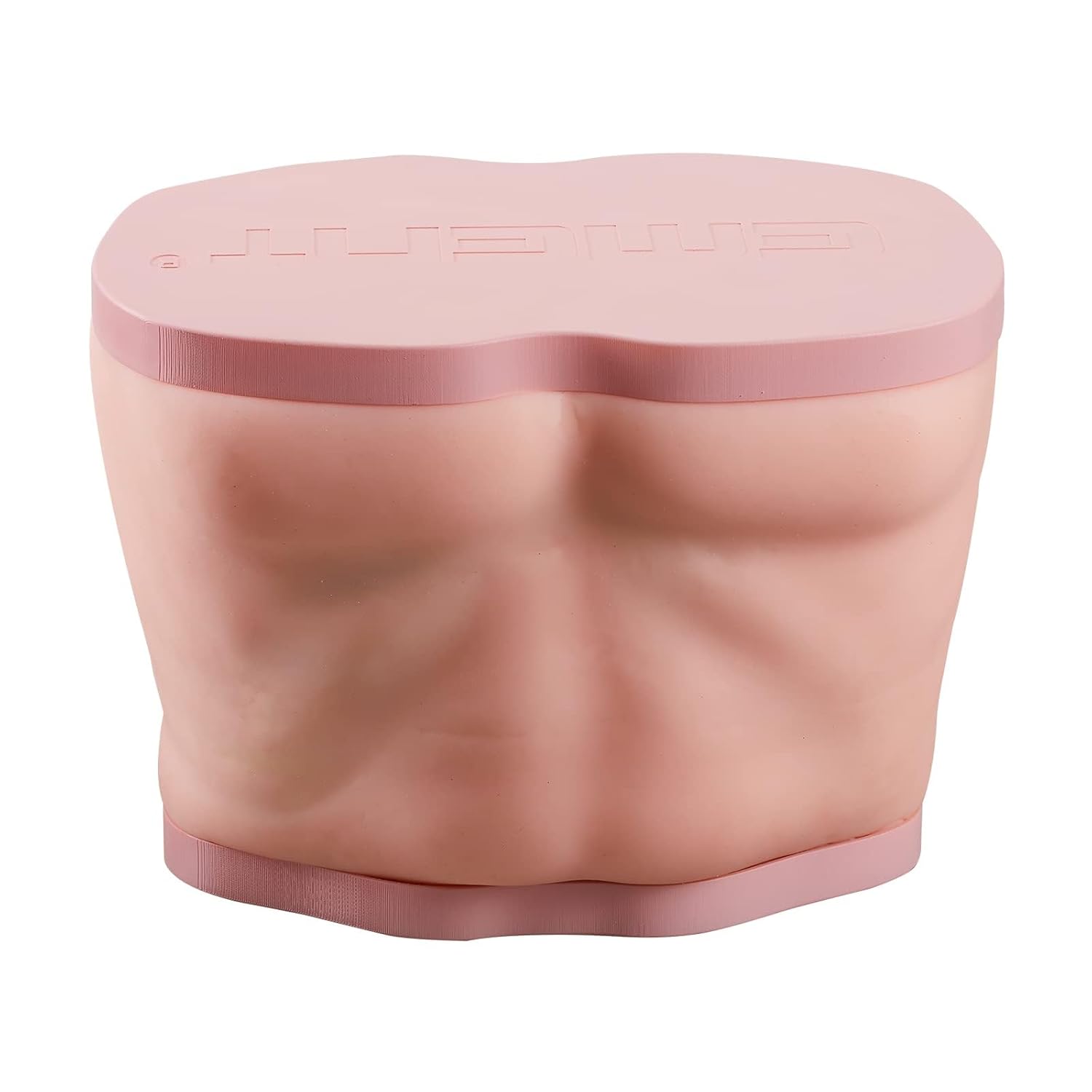
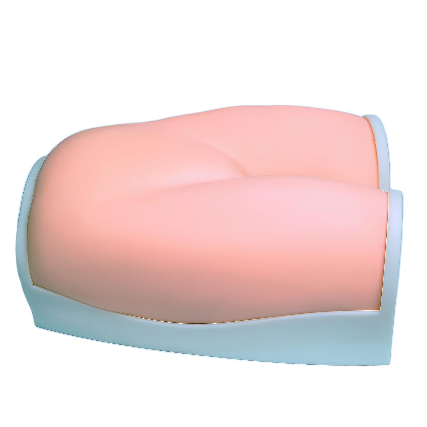
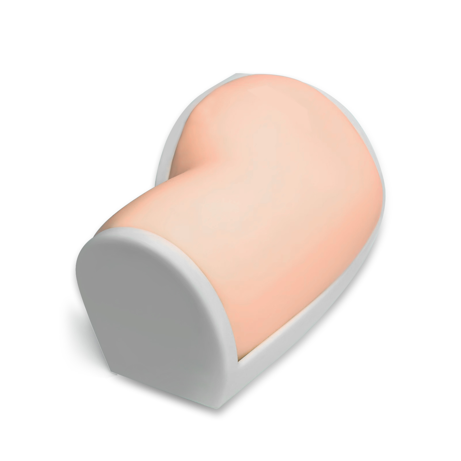
A 1:1 reproduction of the anatomical layers of the radial artery in the forearm, crafted from medical-grade biomimetic acoustic materials. Under ultrasound scanning, vascular borders are distinct, tissue layers are clearly defined, and there is no artifact interference, highly replicating a real clinical ultrasound view. It can be used for core training in ultrasound-guided vascular identification, puncture site localization, and prediction of needle insertion angle and depth, helping operators quickly develop spatial awareness for ultrasound-guided procedures. 
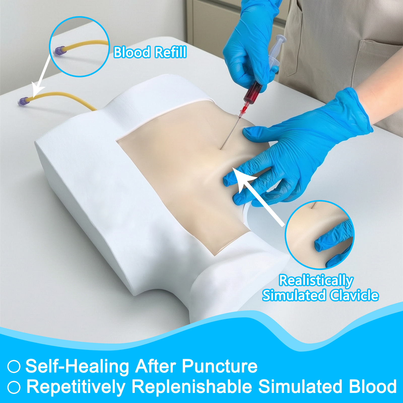
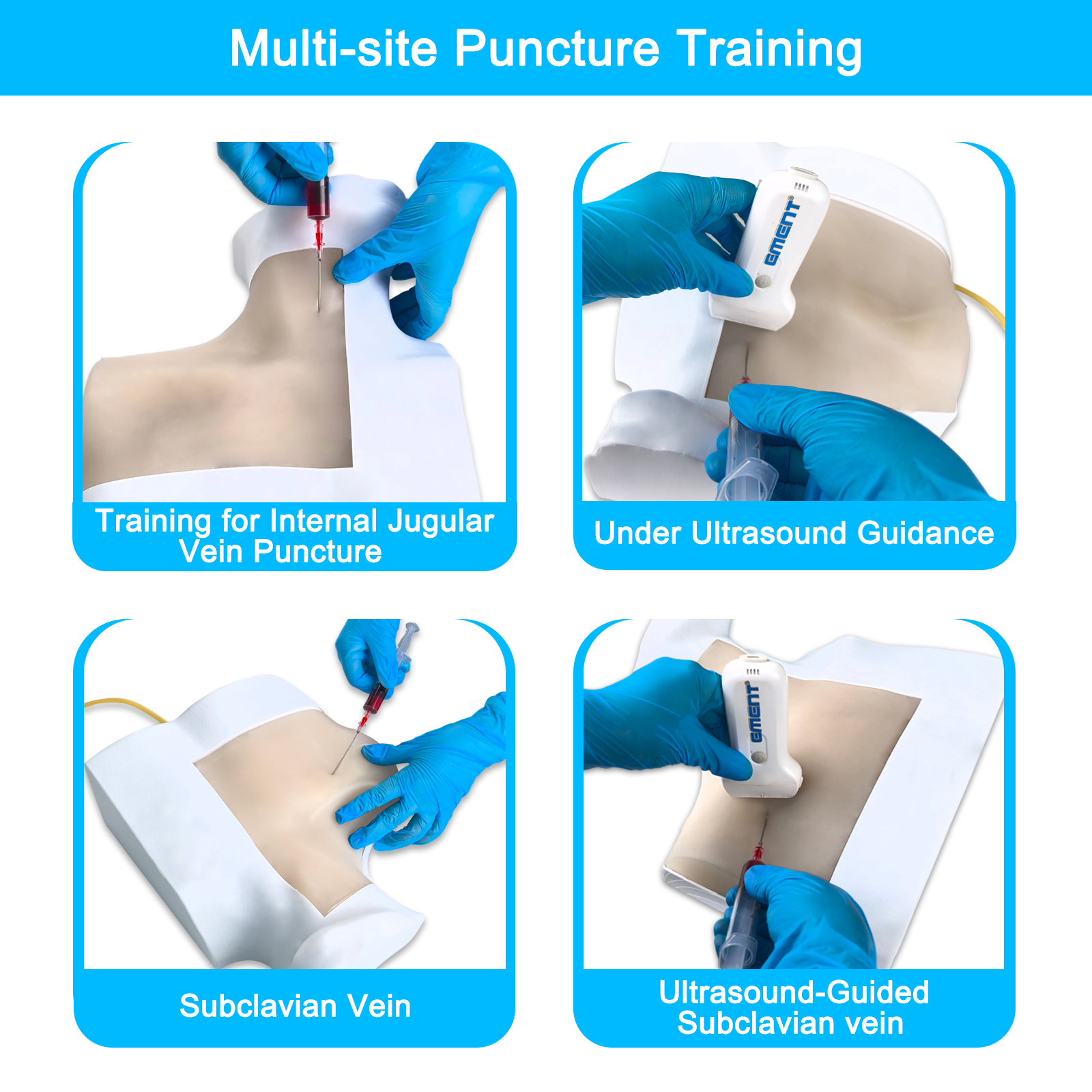
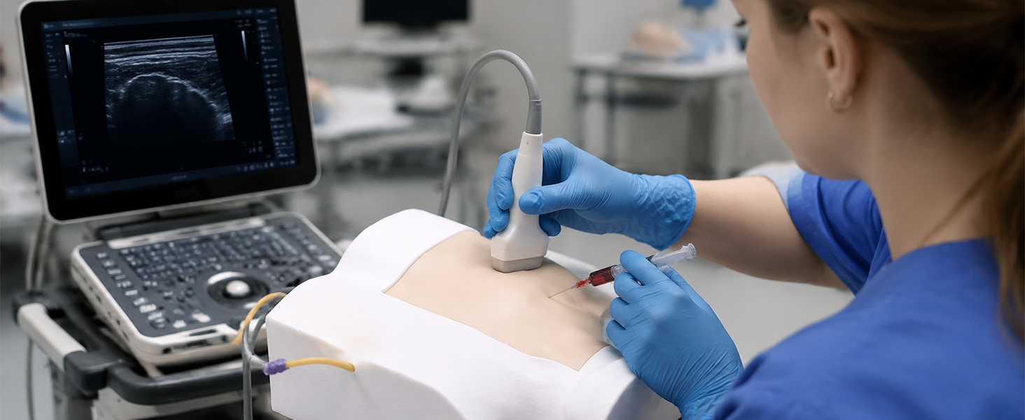
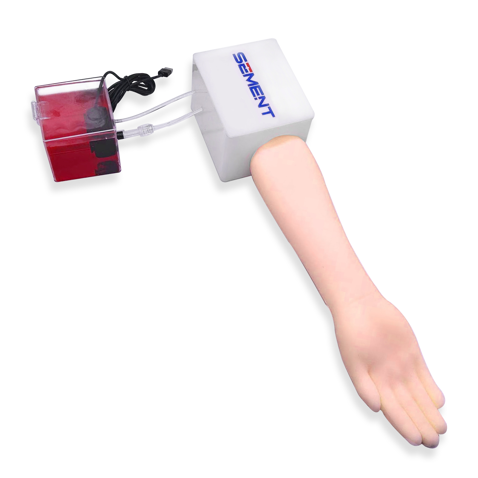
2. Realistic pulsation and backflow feedback, with highly realistic simulation of the entire procedure 
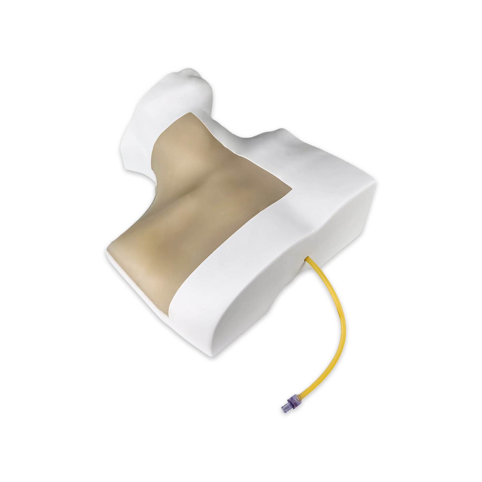
The device features a built-in simulated arterial pulsation system, supporting training for the entire procedure—including ultrasound-guided radial artery puncture and catheterization, as well as arterial blood collection. The simulated skin and blood vessels provide needle insertion resistance and a puncture sensation that closely mimic real-world clinical sensations. Upon successful puncture, clear backflow feedback is displayed, creating a highly immersive training experience that effectively shortens the clinical adaptation period for beginners. 3. Medical-grade, durable material offering excellent value for frequent use 
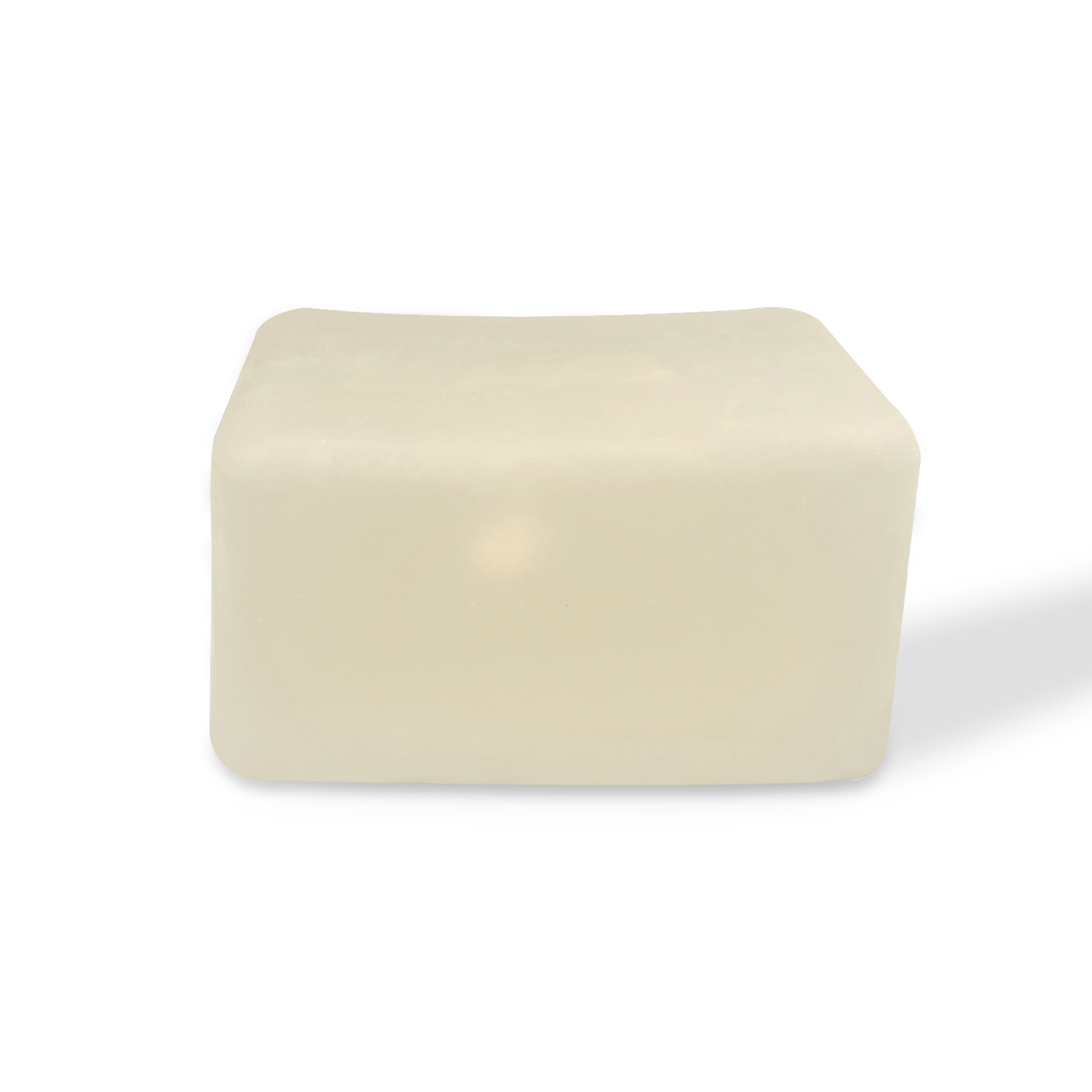
Made from high-density medical-grade simulated silicone, it is safe and odorless. It withstands repeated needle punctures and shows no visible puncture marks even after hundreds of punctures. It resists leakage and deformation, and its ultrasound visibility remains stable without degradation. The smooth surface is easy to clean and can be wiped down with medical-grade disinfectants. It is well-suited for high-frequency training scenarios in medical schools and hospitals, offering a long service life and effectively reducing the cost of training consumables. 
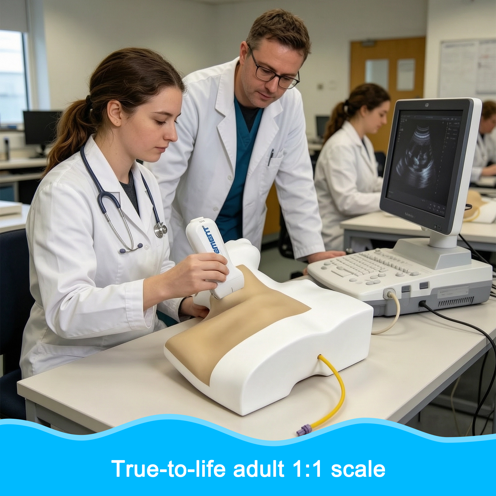
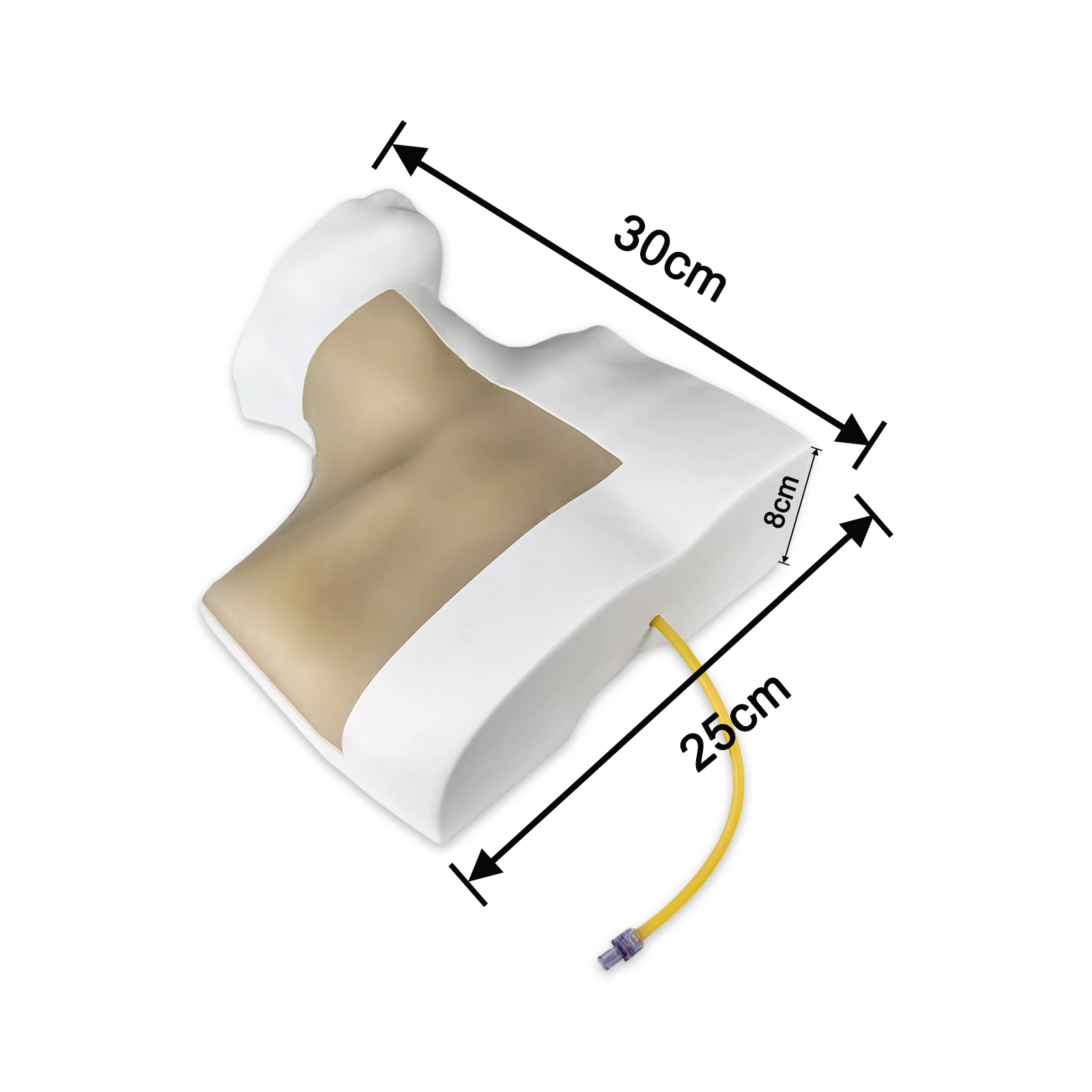
4. User-friendly and highly adaptable design—ready for training right out of the box Featuring an ergonomic, modular design, it stands securely and is easy to operate, requiring no complicated installation or setup. It is compatible with a wide range of commonly used clinical ultrasound diagnostic devices and can be used with standard medical consumables such as conventional puncture needles, indwelling needles, and syringes. It can be flexibly placed on a desk or training table, making it suitable for various training settings, including classrooms, training labs, and clinical departments. 5. A versatile training aid for multiple scenarios and an essential tool for skill enhancement Widely applicable in various settings, including anesthesia, nursing, and emergency medicine education at medical schools; resident training and skill assessments in hospital anesthesia departments, ICUs, and emergency departments; specialized nurse training; and pre-hospital emergency response drills. This is a specialized training aid designed for standardized practice of ultrasound-guided radial artery puncture. It helps beginners quickly master the fundamentals of the procedure and is also suitable for skill reinforcement and assessment among healthcare professionals, effectively reducing risks associated with clinical procedures.

Ultrasound-Guided Testicular Aspiration Training Model

1. High-precision biomimetic anatomical structures for clear and lifelike ultrasound imaging This model faithfully reproduces key anatomical structures and tissue layers of the male testis, epididymis, and spermatic cord on a 1:1 scale. Crafted from medical-grade biomimetic materials, it produces clear and natural images under ultrasound scanning, with distinct tissue layers and no artifact interference, highly replicating a real clinical ultrasound view. It can be used for training in core skills such as anatomical identification, lesion localization, and prediction of needle insertion angle and depth under ultrasound guidance, helping operators quickly develop spatial anatomical awareness during ultrasound procedures. 2. Supports full-process interventional procedures, authentically replicating the clinical feel Supports full-process training for various interventional procedures, including ultrasound-guided testicular biopsy and drug injection; the simulation closely mimics the clinical feel of real human tissue, accurately reproducing the flexibility of the skin and soft tissues, needle insertion resistance, and the sensation of penetrating tissue. This allows users to repeatedly refine their puncture techniques and operational details, effectively shortening the clinical adaptation period for novices and improving procedural accuracy. 3. Made from durable, medical-grade material, offering excellent value for frequent use Made from high-density medical-grade silicone, this product is safe and odor-free. It withstands repeated needle punctures and remains resistant to visible puncture marks, cracking, and leakage even with long-term use. Its ultrasonic visibility remains unaffected by multiple punctures. The smooth surface is easy to clean and can be disinfected with medical-grade disinfectants. Designed for high-frequency use in hospital and academic training settings, it offers a long service life and effectively reduces the cost of training consumables. 4. A versatile training tool for multiple scenarios and an essential tool for skill enhancement Widely applicable in various settings, including urology/andrology education at medical schools, interventional ultrasound training, standardized training and skill assessments for medical professionals, preoperative simulation for clinicians, and specialized training exercises; this is a dedicated training device for standardized training in testicular ultrasound intervention skills. It helps beginners quickly master fundamental techniques while also serving to reinforce skills and conduct assessments for medical professionals, effectively reducing risks associated with clinical procedures.

Ultrasound-guided gallbladder puncture model

A 1:1 scale realistic simulation of an adult liver; used for imaging the intrahepatic bile ducts and common bile duct; simulates the drainage of bile fluid; Four application scenarios: 1. Percutaneous transhepatic cholangiography (PTCD) catheter placement 2. Gallbladder puncture and fistula creation 3. Liver abscess puncture 4. Periliveric fluid drainage

Ultrasound-Guided Oocyte Aspiration Puncture Model

【Realistic Oocyte Retrieval Simulation】The Ultrasound-Guided Oocyte Aspiration Puncture Model is designed for egg retrieval and ovum collection training. It offers a high-fidelity simulation of real-life procedures, making it ideal for practicing oocyte retrieval surgery and ultrasound-guided aspiration techniques in fertility training. 【Hands-On Procedure Training】This trainer is perfect for practice of the egg retrieval process, including oocyte collection and needle insertion under ultrasound guidance. It replicates real-world surgical conditions, allowing users to refine their skills in a safe, controlled environment. 【Anatomically Accurate and Durable】With precise anatomical details, the model provides realistic practice in fertility procedures. The high-quality materials ensure durability, making the model reusable for ongoing training and simulation of retrieval and surgery techniques. 【Ideal for Fertility Specialists and Educators】Perfect for fertility professionals, medical students, and surgical trainers, this model allows hands-on experience in egg retrieval and ovum collection. It’s an essential tool for anyone looking to improve their skills in oocyte retrieval surgery. 【Comprehensive Training Tool】Use this model to practice the entire procedure from ultrasound-guided aspiration to egg retrieval. It is a reliable resource for training, ensuring students and professionals are fully prepared for real-life fertility treatments.

Ultrasound-Guided Abdominal Puncture Training Model

【Abdominal Ultrasound Puncture Training】 6 simulated lumps and 1 Effusionof varying sizes are present inside the tissue in the ultrasound-guided deep tissue mass puncture training model for practice, a unique tool for your practical training and exercises that test your associated skills. 【Excellent ultrasonic guidance performance】It adopts polymer materials equivalent to human tissue, and ultrasonic development is equilalent to human tissue density. Different gray-scale acoustic images are displayed in different tissues when. 【Biopsy puncture and Cyst puncture】 simulate puncture biopsy with different depaths and angles. Cyst treatment, sclerotic simulation, hydrops, pericardial hydrops puncture drainage 【Radiofrequency ablatio,Treatment of hepatic cyst and ovarian cyst】Fistulation of concentrated effusion, one-step, two-part method, PTCD treatment, radiofrequency ablation training. Liver cyst, ovarian cyst treatment, sclerotic simulation, fluid puncture and drainage. 【High Simulation】The size of kidney is th same as that of human body, which truly simulates the ultrasonic image and spatial position distribution of kidney. The cyst model with effusion can be customized.

Ultrasound Liver Puncture Model

【APPLICATION】:Ultrasonic liver puncture biopsy puncture practice training, ultrasound-guided liver tumor puncture biopsy, PTCD placement and drainage, radiofrequency ablation operation, training and teaching demonstration, realistic human skin and animal anatomy. 【PRODUCT INFORMATION】:The ultrasound-guided liver puncture model has a complete contour of the chest and abdomen. The liver is located at the lower edge of the rib cage. There are four tumors in the liver, which are located in different positions of the liver. The ultrasound image of the liver tissue is different from other tissues, so it is easy to identify the position of the liver, and the contour of the image is clear. 【PRODUCT FEATURES AND APPLICATIONS】: Tissue density and liver ultrasound image similar to the liver, there are four masses of different sizes in the liver, distributed in the left lobe of the liver, the middle lobe and the right lobe, complete and beautiful atmospheric shape, is a good partner for ultrasound training, with biopsy needle training, increase the difficulty of training 【Good sound transmission performance】: the material is made of polymer multiple formulations, which can display different gray scale acoustic images in different tissues in order to distinguish between lesions and normal tissues, please use with ultrasound coupling agent and probe, which can be trained with biopsy needles or puncture needles and can be repeatedly used 【After-sales service】: We support a wide range of customized services to suit the needs of every doctor and learner, and for any after-sales problems with the product, our customer service is available 24/7 to answer your questions

Ultrasound Maternity Model

【Witness the Wonders】Our early pregnancy model offers an unprecedented level of precision in depicting the first three months of pregnancy. It provides a clear and vivid representation of various anatomical structures, such as the bladder, uterus, amniotic fluid, and fetal structures. This unique opportunity allows for an up-close examination of the early stages of pregnancy. 【Realistic Simulation】Our pregnancy model is designed with a remarkable level of realism. The material used closely mimics the texture and appearance of real human skin, with an astonishing 95% similarity. This authenticity enhances the learning experience for medical professionals and students alike. 【True-to-Life Uterine Anatomy】In addition to showcasing external changes during pregnancy, our model offers a transparent view of the uterus, revealing its inner structures. This clear visualization allows for a comprehensive understanding of uterine contours and internal organs, providing valuable insights into early pregnancy physiology. 【A Premier Tool for Medical Education】Whether you are a medical student or a healthcare practitioner, our pregnancy model serves as an invaluable educational tool. It offers real-time observation and hands-on experience, deepening your comprehension of early pregnancy physiology. This practical experience is essential for medical professionals in training. 【Preparing for Future Clinical Practice】Our pregnancy model not only enhances your theoretical knowledge but also equips you with practical skills for future clinical practice. It allows for the refinement of medical techniques, from understanding anatomy to practicing delivery procedures. This hands-on experience is crucial for healthcare professionals looking to excel in their clinical careers.Accept customization

Sciatic Nerve Model

【Simulated Sciatic Nerve Model】Our sciatic nerve simulation model provides an unprecedented level of realism for medical professionals and students.Through touch, manipulation, and visual feedback, users can gain a better understanding of the structure and location of the sciatic nerve, preparing them for future medical practices. 【Realistic Image Display】This model is the ideal tool for ultrasound-guided procedure training. It possesses acoustical properties similar to real biological tissues, enabling it to produce clear ultrasound images. Users can employ ultrasound technology to precisely locate the sciatic nerve and perform puncture procedures, enhancing their skill set. This is a valuable training resource for anesthesiologists, nurse anesthetists, and other medical professionals. 【 Applicable Across Multiple Medical Fields】Our model is not limited to anesthesiology but can also be used in neurosurgery, pain management, rehabilitation medicine, and other medical fields.Understanding the anatomy of the sciatic nerve and ultrasound-guided puncture techniques are critical in these areas. 【Can be practiced repeatedly】Adopts polymer material equivalent to human tissue, feel real and no odor.The product is molded in one piece,please use with ultrasound coupling agent and probe.Can be used with biopsy needle or puncture needle training,visible puncture needle path and puncture depth confirmation,can automatically repair the needle channel after puncture. 【Customizable】By gaining a better understanding of sciatic nerve anatomy and puncture techniques, medical professionals can enhance the quality of patient care. You can customize the style to meet your actual practice and training needs.

0
Inquire for more cooperation or product information.
We will contact you within 1 working day, please check your email.
Ultrasound-guided central venous catheterization model
Company Name
Name
Mail
Mobile phone
Message
Send

Yiman

We reply immediately
Welcome to our website. Ask us anything 🎉

Start Chat with: